Procedures
All procedures we offer at NT Cardiac are performed by our cardiologists who have extensive experience in their various fields of expertise. Assisting our cardiologists is our team of highly trained echocardiographers, sonographers and administrative staff.
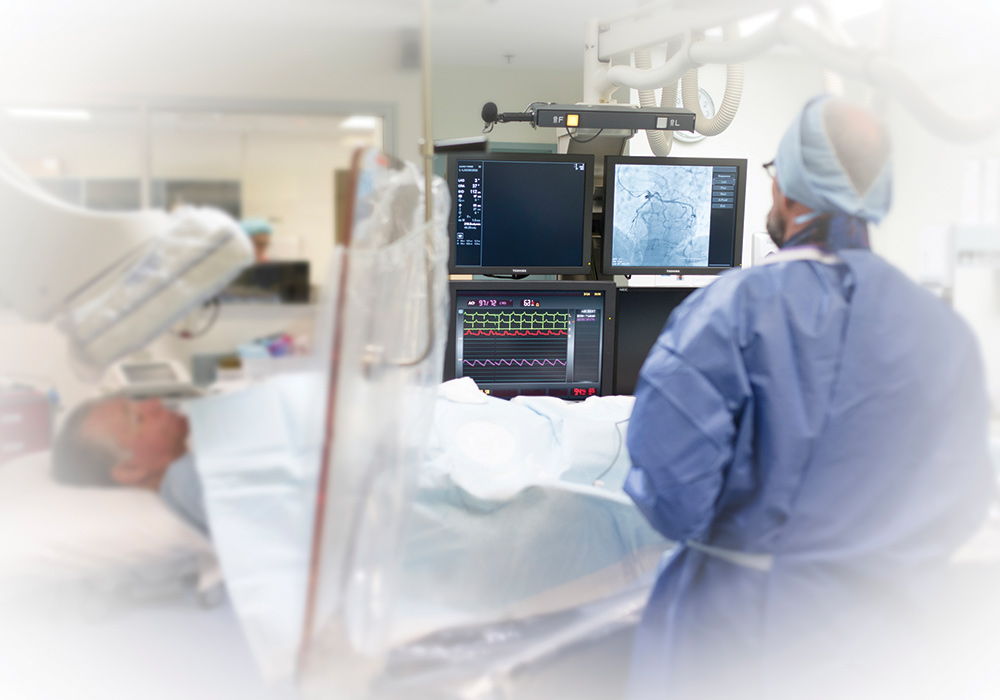
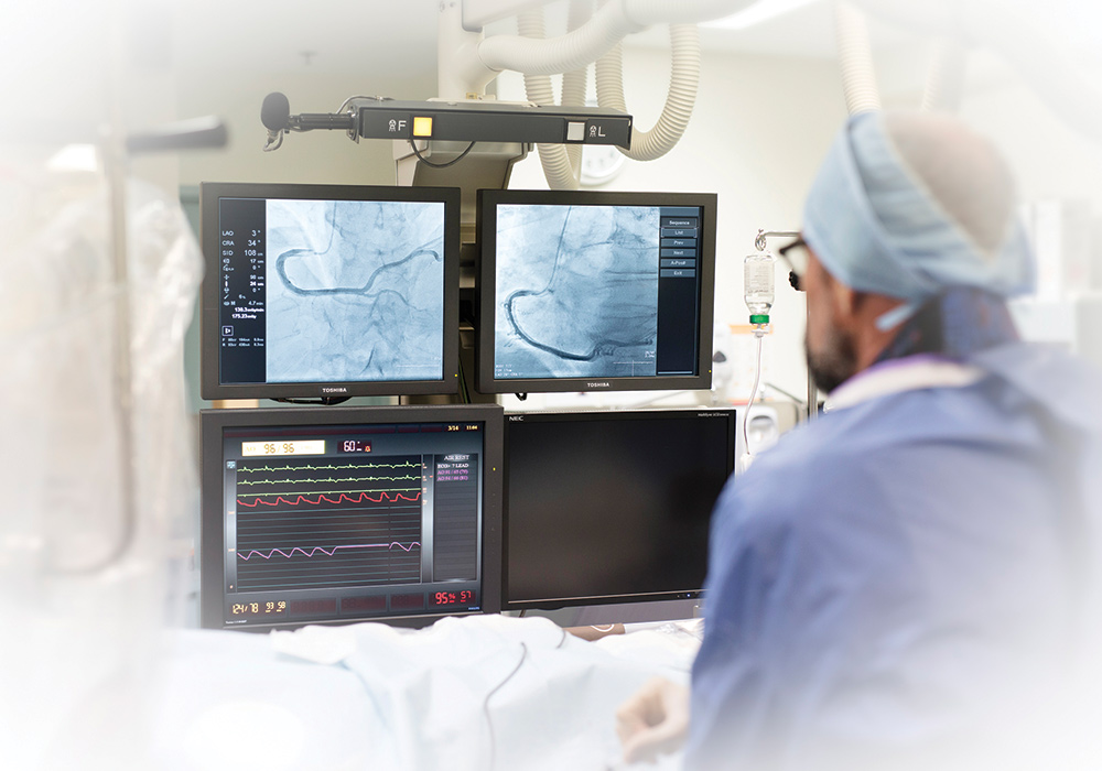
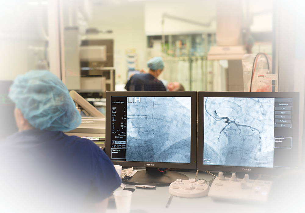
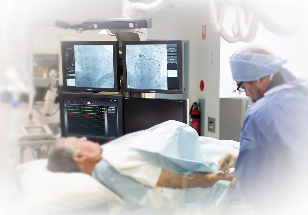
Coronary Angioplasty
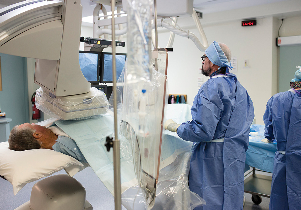
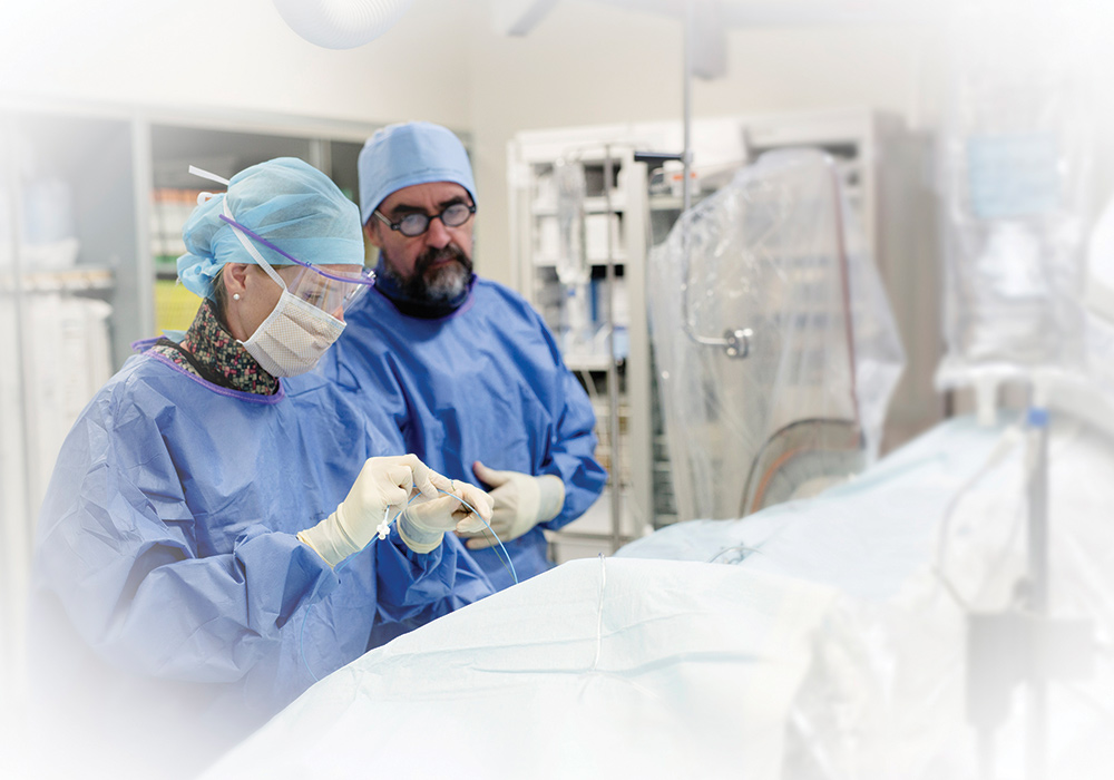
Coronary Angiography is a procedure in which a special X-ray of your heart’s arteries (the coronary arteries) is taken to see if they are narrowed or blocked.
During coronary angiography you are given a local anaesthetic and then a catheter (a long thin tube) is put into an artery in your groin, side of your elbow or near your wrist. The catheter is moved up the inside of your artery until reaches your heart. A special dye is then injected into your coronary arteries and X-rays are taken. The x-rays show the state of your heart and coronary arteries. This is an important test to determine coronary heart disease.
Preparation:
Fast for 4-6 hours prior to procedure.
Duration:
Approximately 40 minutes
Coronary Angiogram
Computed Tomography (CT) Coronary Angiography is a non-invasive scan of the coronary arteries which supply blood to the heart. Through the injections of an iodine contrast via IV cannula and also GTN spray under the tongue.
The coronary arteries and heart chambers are visualised through the injection of a contrast medium during the scan. For optimum results, your heart rate needs to be between 50-60bpm. We may need to give you beta blocker medication to assist in slowing your heart rate down.
The coronary arteries and heart chambers are visualised through the injection of a contrast medium during the scan.
Preparation:
Preparation for the test is longer than the duration of this test.
Patient will asked to fill out the health questionnaire and an ECG will also be taken prior to the test
A 18G IV cannula insertion will also take place mainly on right cubical fossae (on your arm) to prepare for the test.
Duration of test:
Approximately 10 minutes
Cardioversion
Cardioversion is used to return your cardiac rhythm to normal by passing an electric current through the heart muscle. This treatment is mostly used for patients with atrial fibrillation or atrial flutter.
Preparation
If you are on Warfarin you will need to have an INR blood test the day before. Four hours prior to your admission to the cardiac care unit or cardiac surgical unit you will need to fast—no food or drink.
What to bring on the day:
- Medicare card
- Any concession or private health fund cards
- All medication currently being taken, in the original packaging
If you are staying overnight please bring nighttime attire—including dressing gown, slippers and toiletries.
Procedure
A cardiologist, anaesthetist and nursing staff will be present for this procedure.
You will:
- have a small intravenous line inserted into your arm
- be connected to a cardiac monitor
- be given oxygen to breathe
- have a light anaesthetic administered.
You will be asleep when the cardioversion is performed.
Two paddles are used for this procedure. Typically both paddles are placed on your chest; however, there are some cases when one may be placed on the chest and the other on the back.
On waking, you may feel drowsy and we advise you rest in bed until you have recovered from the anaesthetic. It is also important to keep in mind you may, or may not, experience some chest soreness.
Discharge
Depending on how you are feeling you can either leave later the same day or the next morning. A follow-up appointment will be arranged before you leave.
Permanent Pacemaker Insertion
Pacemakers are used to supplement the electrical activity of the heart in patients whose heart is beating too slowly, i.e. “bradycardia”. A pacemaker usually has no benefit for a heart beating too quickly.
A Biventricular pacemaker paces on both sides of the heart. In some cases resynchronising the heart in this fashion can improve cardiac function.
Pacemakers comprise of two components: the generator and the wires.The generator is roughly the same size as two 50c coins and is placed under the skin beneath your collarbone. This is connected to wires which rest inside your heart. These wires are very soft and flexible and can withstand the twisting and bending caused by body movements.
Preparation
You will be required to fast for 6 hours before admission. If you are on blood thinning medication such as Warfarin, you may be required to stop this a few days before the procedure. Your cardiologist will let you know during your consultation.
Procedure
On admission to the hospital, you will have a blood test and ECG.
Your chest will be shaved and painted with Betadine, which is an antiseptic.
Inserting the pacemaker can take anywhere from half an hour to an hour, and is carried out under light sedation—you will be awake but drowsy during the procedure.
Once the area is numb the doctor will make a small cut (approximately 4-7cm) to insert the pacemaker.
The leads are then guided through a vein into your heart and connected to the generator.
Your skin is then stitched together and a small dressing placed over the wound. If the stitches are not dissolvable, they will be removed 7 days after the procedure.
If you require a shower while the dressing is still intact, you will need to cover it in plastic to prevent it from getting wet.
Your Recovery
You will be discharged when your cardiologist is happy with the progress you have made: this can be anywhere from 1 to 3 days post-op.
Before being discharged you will be provided with a detailed booklet on living with a pacemaker.
Following Discharge
- If you have a temperature let your cardiologist know ASAP
- If you are not sure about your medication please contact your doctor
- while the wound is healing, avoid wearing tight clothing over the wound
- If your wound becomes red, swollen or starts to ooze let the hospital know ASAP
- When you shower or bath make sure you keep the wound dry
A follow-up appointment will be organised 2-4 weeks after your procedure.
Automatic Implantable Cardioverter-Defibrillator Insertion
An Automatic Implantable Cardioverter-Defibrillator (AICD) is a small electronic device implanted in your chest and is used to monitor and correct an abnormal heart rhythm, or arrhythmia.
This device is used to treat life-threatening arrhythmias and is the most effective way of doing so.
Defibrillators can treat arrhythmias in two ways:
- tries to stop the abnormal rhythm with a burst of high-speed pacing (cardioversion)
- if that doesn’t work, the device will deliver an electric shock to the heart to reset its rhythm (defibrillation).
Generally speaking, patients aren’t aware of cardioversion but are aware of defibrillation.
The AICD can also collect and store information about your heart’s electrical activity for your cardiologist to check.
Your cardiologist will program the defibrillator to deliver the most appropriate therapy for your condition.
Preparation
You can expect to be in the hospital for 1 to 2 nights. An ECG will be performed, and you may need to have a blood test.
Six hours before admission time you will need to start fasting, and if you on blood thinning medication this may need to be stopped before the procedure.
Procedure
The AICD procedure takes about an hour and is performed under light sedation. An area below the collarbone will be numbed with local anaesthetic and your cardiologist will make a small incision (approximately 5-8cms) to insert the AICD.
Once this is done the leads are then guided through a vein into your heart and connected to the device. The wound is then stitched and a small dressing applied.
When you return to the cardiac unit your heart rhythm and blood pressure will be monitored. Also, you will have an ECG as well as an x-ray.
Following discharge
Post-operatively you will stay in the hospital for 1 to 2 nights and be discharged when your cardiologist is happy with your progress.
Before you are discharged you will be given a booklet with detailed information about living with an AICD.
A follow-up appointment will be organised 2 to 4 weeks after your procedure.
Implanted Loop Recorder Insertion
Have you been experiencing symptoms of recurrent loss of consciousness, palpitations, dizziness, or light-headedness? You might need an implantable loop recorder (ILR).
An ILR is a small device inserted under the skin to monitor and record your heart’s electrical activity over a long period to identify an irregular heart rhythm.
The ILR can determine if your symptoms are related to a heart rhythm problem.
How is the Implanted Loop Recorder fitted?
An Implanted Loop Recorder can be placed in a day and requires you to fast (from food and drink) for 4 hours before the procedure.
A local anaesthetic is used to numb the area and then he ILP is inserted beneath the skin in the upper chest area.
The procedure takes around 20 minutes, and once it has been positioned it can be stitched up.
What happens after the ILR is fitted?
Your cardiologist will program the ILR to the required settings, which only takes a few minutes.
Programming and data retrieval is radiofrequency, and the procedure is virtually painless. Before you are discharged your cardiologist will explain how and when to use your activator.
How do I capture and record an episode?
An ILR captures your heart’s activities during a blackout, which allows your cardiologist to diagnose or rule out an abnormal heart rhythm.
In order to capture and record an episode, you need to place a handheld activator over the ILR. Then press the button to save information.
You should capture the information while experiencing symptoms or as close to the event as possible. The ILR can record what happened 4 minutes before the activator was pressed.
While the ILR can be set to automatically detect an abnormal rhythm without using the activator; it’s still useful to use the activator to show your cardiologist what your heart rhythm is doing when you are feeling unwell.
The device can only store the last 3 activations before it starts to overwrite older recordings.
Removing your ILR
Once your heart activity has been recorded during your symptoms and your cardiologist is satisfied any heart-related causes are identified or ruled out, the device will be removed.
Right Heart Catheterisation
Right Heart Catheterisation is a study of the flow and pressures within the right side of the heart.
This test can be valuable in the assessment of cardiac valvular disease, especially for surgery, and for the assessment of breathlessness.
It involves the insertion of a long thin catheter into a vein in the groin under local anaesthetic. The catheter is moved up the inside of the vein until it reaches the heart. When the catheter is placed, the pressures within the heart can be measured.
A Right Heart Catheterisation is often performed at the same time as a Coronary Angiogram. This procedure can take up to 15 minutes.
Preparation
You will need to fast for 6 hours prior to the procedure, which is carried out in a hospital and is generally carried out in a day. Some patients may be required to stay overnight depending on how they recover.
Take all of your medication as usual unless instructed otherwise.
You cannot drive after the procedure and should not be left alone that night.
Results
You will receive your results at the end of the procedure, and a follow-up appointment will be organised a few weeks afterwards.
These procedures are continuously being developed to improve the management and treatment success of diseases, injuries, and congenital or acquired abnormalities of the heart. For more information, talk to our friendly team.